Sunday, March 27, 2011

The Development of Femtosecond Lasers for Cataract Surgery

Several years ago, in October 2008, I came across a writeup in Cataract & Refractive Surgery Today (CRST), about Technolas Perfect Vision announcing that the newly formed company would be using femtosecond laser technology to investigate laser-based treatments for presbyopia, including the use of intrastromal ablation (ISA). That statement piqued my interest, since I had been involved with the concept of ISA since my earliest involvement with ophthalmic lasers, beginning in the mid-1980s. I decided to write about my engagement with this technology and its history. That became “Intrastromal Ablation: A Technology Whose Time Has Come?

In that article, I wrote about Automated Laser Systems; its successor, Phoenix Laser Systems; another company working on ISA at the time, Intelligent Surgical Lasers; and, finally, the followup of ISL’s work with picosecond lasers that became the laser development work done at the University of Michigan’s Ultrafast Laser Center, that spawned IntraLase, and its femtosecond (FS) laser.

That is where my history of the development of femtosecond lasers stopped – although I did mention the FS lasers being developed by Technolas Perfect Vision. Carl Zeiss Meditec, and Ziemer Ophthalmic Systems, and the then startup LenSx, begun by former Intralase founders. In addition, I also wrote about the work being done by Dr. Luis Ruiz, using the Technolas FS laser in treating presbyopia.

Then, this month, Stephen Daily, news editor for CRST, wrote about what happened to the FS laser companies in the several years after my story. His story, “The Origins of Laser Cataract Surgery: Three companies' pathways from development to commercialization” picks up where my story ended.

With the permission of Stephen and the publishers of CRST, I would like to reproduce his article, to bring the story of the development of femtosecond lasers up to date.


The Origins of Laser Cataract Surgery Three companies' pathways from development to commercialization.

Cataract & Refractive Surgery Today - March 2011

By Stephen Daily, News Editor

This year, the technology behind one of the most anticipated advances in years -- laser cataract surgery – begins its transition from testing laboratories to physicians' offices. The precision of femtosecond lasers in cataract surgery is expected to enhance outcomes in practically all areas of measurement, especially with premium IOLs, which depend on a well-centered capsulotomy. The three major players in the laser cataract surgery market are Alcon, Inc. (Hünenberg, Switzerland), which purchased LenSx Lasers, Inc.; LensAR, Inc. (Winter Park, FL); and OptiMedica Corp. (Santa Clara, CA). These companies plan to commercially launch their devices this year and will go down as the pioneers of the technology. The path each company took to make it to commercialization, however, was unique.

Intralase to LenSx

Similar to many other innovative devices in ophthalmology, femtosecond lasers were originally conceptualized and developed for use unrelated to their potential. The roots of laser cataract surgery can be traced to the work of Ron Kurtz, MD, and Tibor Juhasz, PhD, the founders of IntraLase Corp., who between 1995 and 1997 developed the IntraLase Femtosecond Laser at the University of Michigan in Ann Arbor. The new technology was built for corneal surgery. Knowing the potential to improve LASIK and corneal refractive procedures, Dr. Kurtz and Dr. Juhasz raised $1.4 million in seed money and then approached William Link, PhD, who previously founded American Medical Optics and Chiron Vision and was, at the time, a partner with Brentwood Venture Capital (Los Angeles, CA).

"When I decided to invest, I said to those guys, `I think to do this well, I need you to move to Southern California nearby where we can build a business together,'" Dr. Link said in an interview with Cataract & Refractive Surgery Today.

Dr. Kurtz and Dr. Juhasz made the move to Irvine, California, with the intention to build technology to improve LASIK and corneal refractive procedures, Dr. Link said. During the reduction-to-practice phase, they found in early 2000 that the intrastromal procedure did not work, (emphasis added by I.J. Arons) despite the investment of 5 years and $11.5 million. Dr. Link and the rest of the team decided to refocus their efforts on developing the best device for creating LASIK flaps and later for corneal transplants. They raised $95 million at the initial public offering. Dr. Link attributed the company's ultimate success to "a talented team, substantial capital, and ruthless focus."

In March 2007, IntraLase was acquired by Advanced Medical Optics, Inc., for $808 million in cash. Advanced Medical Optics was acquired by Abbott Laboratories in February 2009, and Abbott Medical Optics Inc. (AMO; Santa Ana, CA) became Abbott's eye care unit. The femtosecond laser developed by IntraLase and now owned by AMO is still trademarked under the name IntraLase.

Before IntraLase was sold to AMO, Dr. Kurtz left the company to pursue the use of femtosecond technology to improve cataract surgery. In 2008, he founded LenSx Lasers, Inc.

The venture capital backers of LenSx include Versant Ventures (Menlo Park, CA), SV Life Sciences (Boston, MA), Interwest Partners (Menlo Park, CA), and Venture Investors (Madison, WI). LenSx received FDA clearance for its laser to create anterior capsulotomies in August 2009, followed by clearance for the creation of corneal incisions in December 2009. In February 2010, Stephen G. Slade, MD, the medical director for LenSx, performed the first laser cataract surgery in the United States on 50 consecutive eyes.(1) Dr. Slade, chief medical editor of CRSToday, said that all of the patients he operated on saw 20/25 or better the first day after surgery, and all of the capsulotomies were perfectly centered and achieved a diametric accuracy of -0.25 mm. In July 2010, Alcon, Inc., announced its purchase of LenSx for a total deal consideration of $744 million, validating the value and interest of the new technology in the ophthalmic marketplace.

LensAR

The pathway to commercial viability for LensAR's femtosecond laser traces back to an original intention of presbyopic correction. Randy Frey was the founder and CEO of Autonomous Technologies Corp. (Orlando, FL), which later merged with Summit Technology (Waltham, MA) and was eventually acquired by Alcon. In 2004, he founded Lasersoft Vision, the predecessor of LensAR, and brought with him some of his partners from Autonomous. Throughout his career, Mr. Frey has been awarded dozens of patents in the area of excimer laser radar tracking, small-beam scanning, and wavefront-guided customized treatments. When he founded Lasersoft Vision, the idea was to research a laser in situ treatment for presbyopic correction, Monty Allen, chief financial officer of LensAR, told CRSToday.

Unlike LenSx, which had venture capital-backed money to help develop and test the technology from the beginning, when Lasersoft Vision started, it was angel funded. Mr. Frey lined up investors who knew of his skills, his capabilities, and his history in ophthalmology, Mr. Allen said. Even Mr. Frey himself was a significant investor in the early stages of the company.

In the course of studying laser techniques for presbyopia, medical advisors at Lasersoft Vision pointed out that the procedure would be ideal for ease of removal of lenticular material as a function of a cataract procedure, especially the lenses that surgeons have the most difficulty breaking up and removing such as higher-grade cataracts.

"In some of the work done in both porcine and human cadaver eyes initially, it was noticed how easily the lens extracted after these treatments, when you needed to extract the lens, for example, to run tests on it," Mr. Allen said. "Some of our physician consultants just simply said we should use this for cataracts. It would make extraction of the lenticular material so much easier, so much faster, and so much less traumatic in terms of the negative side effects from the pounding of the ultrasound energy that causes waves of energy to ripple through the globe. [Potentially] a major cause of anterior segment trauma and endothelial cell loss."

In 2007, after system software upgrades and successful animal trials, Mr. Frey turned his attention to cataract surgery. The company changed its name to LensAR and acquired venture capital. The new focus was to design and develop a highly integrated measurement technology within a three-dimensional scanning laser system. The result was the LensAR Laser System. The primary venture capital partner is Aisling Capital (New York, NY), which was also previously involved as an investment banker for Autonomous Technologies.

The focus on cataracts allowed LensAR to move toward 510(k) clearance, a shorter approval pathway than the full premarket approval the company seeks for its presbyopic indication. An analysis of clinical results with the LensAR Laser System showed a trend toward faster and better anterior capsulotomies with respect to centration and regularity. Also shown were speedy visual recovery, a much reduced use of ultrasound in high-grade cataracts, and the elimination of ultrasound in the lowest-grade nuclei during cataract surgery using laser lens fragmentation versus traditional techniques.(2) In May 2010, the company received FDA 510(k) clearance for use of the LensAR Laser System to create the anterior capsulotomy during cataract surgery. Clearance for laser fragmentation (chop and cylinder patterns) is under active review at the FDA. Other indications are being pursued as well, and the company expects to commercially launch the product in the second half of 2011. (See addendum for further update.)

OptiMedica

The development of femtosecond laser technology at OptiMedica occurred more behind the scenes.

The company was founded in 2004 by five entrepreneurs, including Mark Blumenkranz, MD, chair of ophthalmology at Stanford University. The other founders were George Marcellino, PhD, David Mordaunt, Dan Andersen, and Mike Wiltberger.

Mark Forchette, who has been the president and CEO of OptiMedica since 2007, said there were three areas of focus when the company began-retina, glaucoma, and in the background, in a really "stealthy way," laser cataract surgery.

"We kept it very quiet. Even in the original funding presentation, femtosecond laser cataract surgery was part of it," said Mr. Forchette, who spent 23 years at Alcon before moving to OptiMedica.

Mr. Forchette said the initial investors in the company saw the potential of a precisely controlled capsulotomy and the synergistic effect with the lens that could be there. Those investors include Kleiner Perkins Caufield & Byers (Menlo Park, CA), Alloy Ventures (Palo Alto, CA), DAG Ventures (Palo Alto, CA), and Blackrock Private Equity Partners (Plainsboro, NJ).

The prized possession of OptiMedica up until 2010 was the Pascal Photocoagulator-pattern-scanning laser technology used for retinal surgery. More than a million patients were treated with the Pascal system, according to OptiMedica, and more than 600 systems were sold in 40 markets all over the world.

In August 2010, OptiMedica sold its glaucoma and retina assets, including its proprietary Pascal photocoagulation system, to Topcon Corp. (Tokyo, Japan). The deal allowed OptiMedica to focus exclusively on the continued development and commercialization of its Catalys Precision Laser System. The sale also provided OptiMedica with significant funding for the global market launch of its laser cataract surgery system in 2011.

"The whole time, from the founding of the company until the acquisition last year, we were working on [laser] cataract surgery in the background quietly," Mr. Forchette said. "There's a lot of intellectual property that we filed early that was very forward-thinking, and it was all about image-guidance of femtosecond laser for cataract, capsulotomy, fragmentation, softening, corneal incisions, astigmatic correction, and so those things we've been thinking about since day 1."

Mr. Forchette said the company immediately involved physicians to collaborate with scientists and engineers in the research and development process at OptiMedica. For example, William W. Culbertson, MD, the chair of OptiMedica's Medical Advisory Board, was involved right from the beginning.

The Catalys Precision Laser System combines a femtosecond laser, integrated optical coherence tomography imaging, and OptiMedica's pattern-scanning technology. The platform helped surgeons achieve greater precision during several critical steps of cataract surgery when compared with manual techniques, according to a study published in Science Translational Medicine.(3) The system is not yet approved for sale in the United States, but Mr. Forchette expects it to launch worldwide this year like its competitors.

Technolas Perfect Vision

A fourth company, Technolas Perfect Vision GmbH (Munich, Germany), recently announced its plans to enter the laser cataract surgery market. At the 2010 European Society of Cataract and Refractive Surgeons meeting in Paris, Technolas introduced a customized lens module for cataract surgery. The company's laser, not yet available in the United States, is also able to perform refractive, intrastromal, and therapeutic procedures. (Again, for an update, see the addendum.)


Mr. Allen may be reached at (407) 641-4889; monty.allen@lensar.com

Mr. Forchette may be reached at (408) 850-7488; mforchette@optimedica.com.

Dr. Link may be reached at (949) 729-4500; bill@versantventures.com.

   1. Slade SG.First 50 accommodating IOLs with an image-guided femtosecond laser in cataract surgery.Paper presented at:Refractive Surgery Subspecialty Day,American Academy of Ophthalmology Annual Meeting;October 15,2010; Chicago,IL.
   2. Edwards KH,Frey RW,Naranjo-Tackman R,et al.Clinical outcomes following laser cataract surgery.Invest Ophthalmol Vis Sci.2010;51:5394.
    3. Palanker D,Blumenkranz M,Andersen D,et al.Femtosecond laser-assisted cataract surgery with integrated optical coherence tomography.Sci Transl Med.2010;2(58):58ra85.


Addendum:

Since the above article was written and published, several additional events have occurred:

LensAR

On March 22nd, LensAR, Inc., a developer of next-generation laser technology for refractive cataract surgery, announced that it had received 510(k) clearance from the FDA for use of the LensAR Laser System for anterior capsulotomy and lens fragmentation during cataract surgery.

“Receiving the additional FDA indication for lens fragmentation is a significant milestone achievement in getting our technology one step closer to commercialization. We are very pleased with the exceptional fragmentation data that was submitted to obtain the indication and the resulting FDA clearance,” said Randy Frey, founder and Chief Executive Officer of LensAR.

B&L and Technolas Perfect Vision

On March 25, 2011, Bausch & Lomb and Technolas Perfect Vision announced an agreement in principle to distribute the first femtosecond laser capable of performing both cataract and refractive surgery on a single platform

"Femtosecond laser technology for cataract procedures promises to be one of the most significant clinical advances in cataract surgery in 40 years," said Robert E. Grant, chief executive officer and president of Bausch & Lomb Surgical. "The TPV femtosecond laser platform, which uniquely supports refractive and cataract procedures, is a natural complement to our portfolio of cataract products. This is the first of many new technologies we are pursuing focused on enhancing a physician's ability to improve patient outcomes."

Commercialization is expected to begin in the second half of 2011. TPV previously announced that they had filed for 510(k) clearance in the United States.

Richard Lindstrom @ ASCRS

On March 26th, Richard L. Lindstrom, MD, spoke at Glaucoma Day preceding the American Society of Cataract and Refractive Surgery meeting in the Cataract/Cornea Crossover Topic. He said femtosecond laser platforms are clinically well-established for LASIK flap creation on five platforms.

"Advances will come in both the imaging and diagnostics technology that would allow us to have applications to other fields, and I firmly believe glaucoma will be one of those," he said. "We will have to sort out, certainly, and understand better the demand and the business model, but I firmly believe that the 78 million baby boomers will probably find this very, very attractive. And the early surgeons who have adopted this technology are not finding much difficulty generating significant interest from their patients as to this alternative, if you will, to manual surgery. So lots more to learn."

For laser refractive cataract surgery, four companies are developing platforms: Alcon LenSx, LensAR, OptiMedica and Technolas Perfect Vision. In addition, three companies, Abbott Medical Optics, Schwind and Carl Zeiss Meditec, are in undisclosed stages of development. The technology offers excellent precision, smaller incisions and enhanced IOL performance, Dr. Lindstrom said. It creates a safer, more predictable and reproducible procedure.


Editor’s Note:


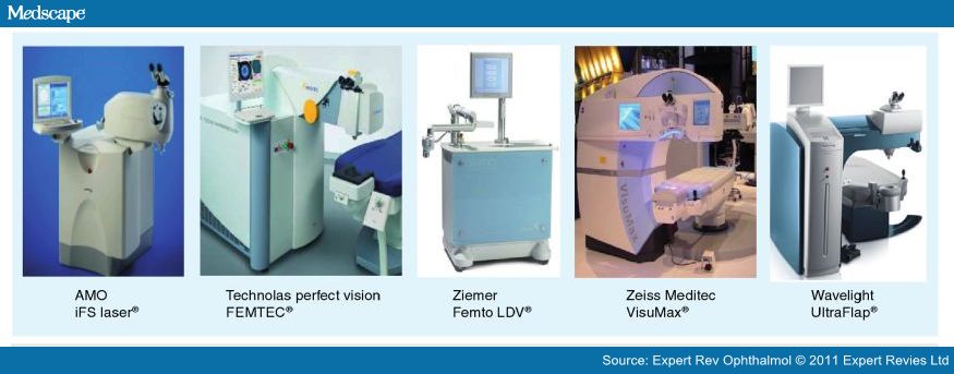
In addition to the history of intrastromal ablation and the introduction of femtosecond lasers article mentioned in the Preface, I have posted four other articles dealing with femtosecond lasers: one describing the FLEx (Femtosecond Lenticle Extraction) method of correcting corneal error, using the Carl Zeiss Meditec VisuMax femtosecond laser (Another Approach to Intrastromal Ablation); two articles about using the femtosecond laser for cataract removal (Femtosecond Lasers Proposed for Use in Cataract Surgery and, Femtosecond Laser Cataract Removal: The Second Revolution? And, What is Laser Photolysis? – the latter describing the possibility of using FS lasers to bleach a cataractous lens to postpone the need for cataract surgery); and, finally, A Comparison of Commercially Available Femtosecond Lasers in Refractive Surgery – which compares the IntraLase IFS (Abbot), Femtec (Technolas), VisuMax (Carl Zeiss Meditec), LDV (Ziemer) and UltraFlap FS 200 (WaveLight/Alcon) systems.

Tuesday, March 22, 2011

Menu 18: Updates for October 2010 – March 2011

I have been negligent in updating the menus for my online Journal. So, here is the update for the past six months.

This update contains a number of new writeups, including the first use of gene therapy in treating retinitis pigmentosa; an overview of what happened at the Second Ophthalmic Innovation Symposium (held last year just prior to the AAO Meeting), a couple of interviews conducted by my colleague in China during lat year’s APAO Meeting in Beijing, and an excellent writeup about how femtosecond lasers are being used in ophthalmology.

I have also included several updates on subjects I have been closely following, including the controversy between the use of Avastin or Lucentis in treating AMD; an AMD update; a CATT Study update; five updates on the use of stem cells in ophthalmology; and updates on the latest news from Iluvien and NeoVista.

Here are synopses and links for the recent postings:

Avastin/Lucentis Updates:
Avastin/Lucentis Update 42: One-Year Results of Controlled Comparison Study Published (Oct. 4, 2010)

Last October (2009), I came across the six-month results of one of the first blinded, double-masked comparison studies run between Avastin and Lucentis, sort of a mini-CATT Study. This study was done by researchers at the Boston University School of Medicine in cooperation with the VA Boston, and was published in the American Journal of Ophthalmology. I published their news release describing the study and the six-month results as Avastin/Lucentis Update 29.

This weekend, the same group announced the one-year results of this study, this time published in Eye, a peer-reviewed publication of The Royal College of Ophthalmologists in the UK.


Avastin/Lucentis Update 43: Secret Rebates Offered for Lucentis (Nov. 4, 2010)

First, Genentech refused to provide Lucentis for the CATT Study, being run by NEI/NIH to compare Avastin to Lucentis for AMD (Avastin/Lucentis Update 12); then they threatened to stop supplying Avastin to compounding pharmacies so that ophthalmologists could continue to obtain the drug for their patients (Avastin/Lucentis Update 18); then they decided to provide Lucentis free of charge for the study looking at the use of panretinal laser treatment plus anti-VEGF (Lucentis) in the treatment of diabetic macular edema – at the exclusion of Avastin, in that “pay to play” study (Avastin/ Lucentis Update 37); and now, the company is offering secret rebates to selected large users of Lucentis – obviously to blunt the potential expected to be offered by Avastin when the CATT Study results are released next Spring.

Here, as written by Andrew Pollack online yesterday and published in today’s NYTimes, is the latest story in the ongoing Avastin vs. Lucentis Controversy:


Avastin/Lucentis Update 44: United Kingdom Closer to Allowing Avastin for AMD (Dec. 8, 2010)

While the U.S. comes closer to showing the equivalency of Avastin to Lucentis for treating the wet form of age-related macular degeneration, when the CATT (Comparisons of Age-Related Macular Degeneration Treatments Trials) Study results become public, hopefully, some time this Spring, the UK’s health services are still fighting over whether or not they should study the two drugs to determine if Avastin would be appropriate for the Brits to use in their National Health Service.


Avastin/Lucentis Update 45: Avastin Drug Treatment for ROP Better than Laser (Feb. 26, 2011)

A new study, published earlier this month in the New England Journal of Medicine, describes the use of intravitreal Avastin to treat Retinopathy of Prematurity (ROP) in premature infants.

This study was widely covered by the press, but I would like to reproduce just a few of the presentations to provide you with the information necessary to best understand the results of this study.


AMD Update:

AMD Update 13: Retinal Procedures on the Rise (Oct. 13, 2010)

A new study, just published in the October issue of Archives of Ophthalmology and reported by Ophthalmology Web, MedPage Today, and Medscape Medical News,  shows that among those in the Medicare population (age 65 plus), treatments for retinal conditions nearly doubled between 1997 and 2007 – and this trend is expected to continue with the aging of the population and seniors living longer.


CATT Study Update:
CATT Study Update 12: Status of WorldWide Studies (Jan. 6, 2011)

With the anticipated arrival of the one-year results of the CATT Study this Spring, I thought it would be appropriate to update where the other worldwide studies stand.

During the Retina Subspecialty Day sessions, held prior to the recent 2010 AAO Meeting in Chicago, Daniel Martin, MD provided an update on the various comparative studies underway around the world between Avastin and Lucentis. Here are Dr. Martin’s comments, as reported by the Market Scope team in the November issue of Ophthalmic Market Perspectives.


Stem Cells in Ophthalmology Updates:
Stem Cells in Ophthalmology Update 2: ACT Gets Go-Ahead to Treat Stargardt’s (Nov. 23, 2010)

As I noted in my September report on the Use of Stem Cells in Ophthalmology, it was anticipated that either the program at The London Project to Cure Blindness or Advanced Cell Technology’s program to treat Stargardt’s disease would be the first to get the go-ahead to begin approved human trials. I have not heard any news out of London, but earlier this week ACT received notification from the FDA that it was cleared to begin its human trials with human embryonic stem cells.


Stem Cells in Ophthalmology Update 3: ACT Files IND to Treat Dry AMD (Nov. 30, 2010)

Furthering its lead in stem cell research in ophthalmology, Advanced Cell Technology Inc., announced today that it  had filed an Investigational New Drug (IND) application with the U.S. Food and Drug Administration, to initiate a Phase I/II multicenter study for the  treatment of dry Age-Related Macular Degeneration (dry AMD) using human embryonic stem cell (hESC) derived retinal pigment epithelial (RPE) cells.


Stem Cells in Ophthalmology Update 4: ACT Receives Receives FDA Approval to Use hESCs to Treat Dry AMD (Jan. 3, 2011)

Advanced Cell Technology Inc., announced today that it  had received approval from the FDA to commence its clinical trial using retinal pigment epithelial (RPE) cells derived from human embryonic stem cells (hESCs) to treat the dry form of age-related macular degeneration. ACT is now permitted to initiate a Phase I/II multicenter clinical trial to treat patients with dry AMD, the most common form of macular degeneration in the world. There are currently no approved treatments available for this prevalent disease of an aging global population. Dry AMD, representing a substantial global market opportunity and afflicts between 10-15 million Americans, and a further 10 million Europeans.

Stem Cells in Ophthalmology Update 5: Gene Defects Common in Induced Stem Cells (Mar. 5, 2011)

As the senior editor, John Gever, of MedPage Today reported, following the publication of three studies about induced pluripotent stem cells in the March 3rd, issue of Nature, “The road to regenerative medicine based on induced pluripotent stem cells (iPSCs) may have developed a giant pothole, with new studies showing that the cells are prone to several types of genetic defects.”

The three studies showed that the reprogramming process and subsequent culture of pluripotent stem cells in vitro can induce genetic and epigenetic abnormalities in these cells. The authors of the studies and the editorialist said that the results raise concerns over the implications of such aberrations for future applications of pluripotent stem cells.


Stem Cells in Ophthalmology Update 6: Stemedica Paper Accepted for Presentation at ARVO (Mar. 19, 2011)

I recently received an update from my contact at Stemedica and would like to share the information with you.

A safety study on the use of  stem cells in the eye, in a clinical study underway at the Fyodorov Federal Institution of Eye Microsurgery in Moscow, to treat diabetic retinopathy and diabetic optical neuropathy with stem cells derived from bone marrow, has been accepted for presentation as a poster at the upcoming ARVO Annual Meeting in Fort Lauderdale at the beginning of May. An abstract is shown below.


Iluvien Update:

Iluvien Update: FDA Marketing Approval Delayed (Jan. 5, 2011)

Last July, I wrote a comprehensive report about Iluvien and the status and promise of other sustained release drug delivery systems (Iluvien and the Future of Ophthalmic Drug Delivery Systems). At that time, Alimera Sciences, the company developing Iluvien (under license from pSivida) had filed a new drug application (NDA) to treat diabetic macula edema (DME). The company obtained priority review status for the NDA at the end of August, raising the expectation that an approvable letter might be obtained by the end of 2010.

However, instead of an approvable letter, Alimera Sciences received a “complete response letter” (CRL) from the FDA, communicating to the company that its NDA application “cannot be approved in its present form”.


NeoVista Update:

NeoVista Epi-Retinal Strontium 90 Treatment for AMD: Update 4 (Jan. 20, 2011)

NeoVista just released an update, discussing the first commercial utilization of its Epimacular Brachytherapy device in Germany. The Epi-Rad device, now renamed as the VIDION ANV (Anti Neo Vascular Therapy System) has been commercialized in Europe since November 2009. The first patients treated were in Pisa, Italy, quickly followed by patients treated in London, UK, also in November of 2009, and now in Hamburg, Germany this month.


And, here are the new writeups:
The Use of Gene Therapy in Treating Retinitis Pigmentosa and Dry AMD by Retrosense (Nov. 6, 2010)

A short while ago, I received a message from someone I did not know, who said that he enjoyed the writeups on my online Journal and was wondering if I might be interested in writing about the use of gene therapy as an approach to vision restoration. Since I knew absolutely nothing about gene therapy, the writer got my attention.

After several discussions with Sean Ainsworth, the founder of RetroSense, and much online research, I think I have learned a little about what gene therapy is about, and its application in ophthalmology, especially in the possible restoration of vision in those who suffer from retinitis pigmentosa (RP). Thanks to Sean for whetting my appetite -- here is what I have learned.


The Second Ophthalmic Innovation Summit (Dec. 10, 2010)

Last October, the Second Ophthalmic Innovation Summit (OIS) was held just prior to the 2010 AAO Meeting in Chicago. As was the case last year, my old friend, Larry Haimovitch,an ophthalmic industry veteran, who attended the meeting and wrote about it for the sponsor’s website, Healthcare Syndicate has given me permission to reproduce his writeup in this space.

An Interview with Dr Ronald Krueger (Feb. 17, 2011)

With the permission of Lei Zang, the Managing Editor of Ophthalmology World Report, here is her interview with noted refractive surgeon Dr. Ronald Krueger of the Cleveland Clinic. As noted below, Dr. Krueger was recently in China to perform a corneal transplant on a Mongolian patient and while there, attended the APAO Meeting held last fall in Beijing, where Ms. Zang had the opportunity to interview him.


An Interview with Professor John Marshall (Feb. 17, 2011)   

During the APAO (Asia Pacific Academy of Ophthalmology) Meeting held in Beijing in September, 2010, Lei Zang, the Managing Editor of Ophthalmology World Report,  interviewed Professor John Marshall of St. Thomas Hospital in London and the principal behind the Ellex 2RT (retinal regeneration) program for Ellex Laser, and also an investigator (and inventor?) of the Avedro microwave corneal crosslinking program.

In this interview, Prof. Marshall discussed both his work on 2RT and on microwave crosslinking as well as several other topics that will be of interest to ophthalmic researchers.

              
A Comparison of Commercially Available Femtosecond Lasers for Refractive Surgery (Mar. 17, 2011)

Over the past several years, I have either written or posted other peoples accounts of the use of femtosecond lasers in ophthalmology. Starting with the history of their development in October 2008 (Intrastromal Ablation: A Technology Whose Time Has Come?); an article on the use of the femtosecond laser to treat presbyopia by Dr. Rupal Shah in June 2009  (Another Approach to Intrastromal Ablation); a writeup on the use of femtosecond lasers for performing cataract surgery by Larry Haimovitch, again in June 2009 (Femtosecond Lasers Proposed for Use  in Cataract Surgery); and finally, Dr. Joseph Colin’s writeup about femtosecond laser cataract removal as a second revolution, and my addition about the possibility of using the femtosecond laser to “bleach” the natural lens to delay the onset of needing to remove catatacts, inAugust 2010 (Femtosecond Laser Cataract Removal: The Second Revolution? And, What is Laser Photolysis?).

Earlier this month, I came across an article written by Drs. Ronald Krueger and Glauco Reggiani-Mello, of the Cleveland Clinic, that does an excellent job of summarizing the latest developments in the use of femtosecond lasers in refractive surgery and other applications in ophthalmology. Since the article was written in a professional journal – Expert Review of Ophthalmology, with limited access, I asked the authors for permission to reproduce a significant part of their writeup, along with a link to the original for those that wish to read it in its entirety. Permission was granted, and here is my version of what was presented, along with most of their illustrations and their two tables.

Saturday, March 19, 2011

Stem Cells in Ophthalmology Update 6: Stemedica Paper Accepted for Presentation at ARVO

I recently received an update from my contact at Stemedica and would like to share the information with you.

A safety study on the use of  stem cells in the eye, in a clinical study underway at the Fyodorov Federal Institution of Eye Microsurgery in Moscow, to treat diabetic retinopathy and diabetic optical neuropathy with stem cells derived from bone marrow, has been accepted for presentation as a poster at the upcoming ARVO Annual Meeting in Fort Lauderdale at the beginning of May. An abstract is shown below.

In addition, my contact told me that Stemedica has registered in Clinical Trials, to conduct a Phase I/II multicenter trial under the auspices of UC San Diego, using bone marrow derived stem cells on patients with post ischemic strokes. The next step for the company will be to get a “green light” from the FDA to treat patients in the U.S., using the same type of cells on patients with retinal disorders. Those trials will include studies both with and without the use of a laser, as explained in my report on the company in the Primer on the Use of Stem Cells in Ophthalmology.


Phase I Study: Injection of Allogeneic Bone Marrow Derived Mesenchymal Stem Cells in Patients with Diabetic Retinopathy and Diabetic Optical Neuropathy

Author Block:
Natalia Gavrilova (1), Khristo Takhchidi (1), Irina Saburina (2), Nikolai Mironov (3), Marina Polyakova (1), Alexei Lukashev (4), Paul Tornambe (5).

(1) Ophthalmology, Fyodorov Federal Institution 'Eye Microsurgery', Moscow, Russian Federation; (2) Institute of General Pathology, Moscow, Russian Federation;
(3) Moscow Neurology Hospital, Moscow, Russian Federation;
(4) Stemedica Cell Technologies, San Diego, CA;
(5) Retina Consultants, San Diego, CA.

Abstract:

Purpose: Diabetic retinopathy (DP) and diabetic optical neuropathy (DON) are common complications of diabetes which are caused by abnormal development of retinal microvasculature. Initial case studies have been performed on human patients to assess the safety and efficacy of this type of cell therapy.

Methods: Six patients (4 female, 2 male, age 42-62) with early stages of diabetic retinopathy and diabetic optical neuropathy were injected intravenously with a single dose of 100M allogeneic bone marrow derived mesenchymal stem cells (BM MSC). The cells were manufactured under the guidelines of current good manufacturing practice (cGMP). All patients were examined prior to the injection and post injection on day 1, 14, 30, 2 month, 3 month, 6 month, one, two and three years. General examination included blood and urine panel, blood coagulation test and measurements of brain derived neurotrophic factor (BDNF) in blood plasma and tear fluid. Ophthalmic examination included visual acuity and ophtalmoscopic exam, perimetry test, optical coherent tomography(OCT), eletroretinography(ERG), electrooculography(EOG), Doppler ultrasound exam, optic disk color analysis.

Results: No adverse effects were observed or reported by all patients. During the whole period of follow up there were no changes in cardiovascular system, liver and kidney functionality. No infection growth, interstitial pneumonia or cancer was found in all patients. The positive changes in hemostatic dysfunctions and well as improvements in hemodynamic at systemic level were observed as early as two weeks after injection. Fovea sensitivity; functional activity of optic nerve, pigmented epithelium layer, outer and inner nuclear layers; BDNF content in blood plasma and tear fluids as well as optic disc inflammation were gradually improved during the first six months post injection and became stable during the whole period of follow up.

Conclusions: The results show safety of intravenous injection of BM MSC on patients with diabetic retinopathy and diabetic optical neuropathy. Positive changes of ophthalmic parameters should be further investigated in full scale clinical trails.

Thursday, March 17, 2011

A Comparison of Commercially Available Femtosecond Lasers in Refractive Surgery

Over the past several years, I have either written or posted other peoples accounts of the use of femtosecond lasers in ophthalmology. Starting with the history of their development in October 2008 (Intrastromal Ablation: A Technology Whose Time Has Come?); an article on the use of the femtosecond laser to treat presbyopia by Dr. Rupal Shah in June 2009  (Another Approach to Intrastromal Ablation); a writeup on the use of femtosecond lasers for performing cataract surgery by Larry Haimovitch, again in June 2009 (Femtosecond Lasers Proposed for Use  in Cataract Surgery); and finally, Dr. Joseph Colin’s writeup about femtosecond laser cataract removal as a second revolution, and my addition about the possibility of using the femtosecond laser to “bleach” the natural lens to delay the onset of needing to remove catatacts, inAugust 2010 (Femtosecond Laser Cataract Removal: The Second Revolution? And, What is Laser Photolysis?).

Earlier this month, I came across an article written by Drs. Ronald Krueger and Glauco Reggiani-Mello, that does an excellent job of summarizing the latest developments in the use of femtosecond lasers in refractive surgery and other applications in ophthalmology. Since the article was written in a professional journal – Expert Review of Ophthalmology, I asked the authors for permission to reproduce a significant part of their writeup, along with a link to the original for those that wish to read it in its entirety. Permission was granted, and here is my version of what was presented, along with most of their illustrations and their two tables.


Comparison of Commercially Available Femtosecond Lasers in Refractive Surgery
Glauco Reggiani-Mello and Ronald R Krueger

Cole Eye Institute - Cleveland Clinic Foundation, 668 Euclid Avenue, Unit 506, Cleveland, OH 44114, USA

Published in: Expert Review of Ophthalmology
Posted online: 03/01/2011

Abstract:


Refractive surgery is a procedure that requires excellence. Nothing less than the best is acceptable for an elective procedure that must be precise, accurate and safe. Femtosecond lasers were developed to help fulfill these requirements and have changed the field. The capabilities of the technology include not only the creation of corneal flaps for laser-assisted in situ keratomileusis, but limitless corneal- and lens-based incisions, as well as glaucoma and retinal applications that can break old paradigms. Manipulating biomechanics to correct presbyopia with the IntraCor procedure, the 'femtosecond-only' femtosecond lenticule extraction or SmILE procedures, intrastromal astigmatic incisions and cataract surgery are among the next exciting applications to this technology. New fields in refractive surgery can be opened and others can be expanded, as in refractive lens exchange, where its indications may be greatly increased, considering the new safety and precision standards that the technology can deliver. The expectations are huge and future studies will show how far we can go with the technology.

Introduction:

Femtosecond lasers have changed refractive surgery in the last 9 years since the market release of the Intralase Femtosecond Laser (Abbott Medical Optics, IL, USA) in 2001. The bladeless flap creation rapidly gained popularity because of its promised increased safety, fast recovery and excellent results.[1] Nowadays, the majority of high-volume refractive surgery centers in the world uses a femtosecond laser to create the flap.

Technical Parameters

All commercially available devices use a near infrared femtosecond laser with a wavelength of approximately 1053 nm. Despite the fact that the neodymium-doped yttrium aluminum garnet and femtosecond lasers have very similar wavelengths (Table 1), the ultrashort duration of the pulses (10-9 vs 10-15) in the latter causes significantly less damage in the collateral tissue.[2] Varying the duration of the laser pulses and energy applied can generate different effects on the tissue (Figure 1). The main technical specifications that play a role in the femtosecond laser are the following:

    * Laser pulse repetition rate;
    * Spot size;
    * Pulse energy;
    * Pulse pattern.


    Figure 1.  Effects of the relationship between laser interaction time and energy intensity.


There is an inverse relationship between the laser pulse duration and the energy required in each pulse to generate the optical breakdown.[3] A shorter pulse (200-500 fs) needs lower energy to achieve the threshold of photodisruption than a longer pulse (500-1300 fs). The numerical aperture (NA) of the lens influences the laser spot in terms of diameter and volume. A higher NA focuses the beam with less dispersion and is the reason why higher NA devices use lower energy. It is also suggested that a higher NA increases the depth accuracy and overall precision of the lamellar cut.

After the optical breakdown occurs, plasma is created and a cavitation bubble formed. This bubble expands and cleaves the tissue. If a high-energy photodisruption is used, the bubble is larger and the pulses do not need to be placed close together. Low-energy systems create a very small bubble, with a greater number of pulses in an overlapping pattern being mandatory, since there is almost no tissue cleaving induced by the bubbles.

The first devices operated with a low KHz repetition rate (15 KHz - first Intralase model) and needed a higher energy to photodissection. Newer devices (even the newer high-energy devices such as IntraLase 150 KHz) intend to increase the repetition rate, which makes the procedure duration shorter and uses lower energy with an intention of diminishing the inflammation. In addition, the spot size and separation can be lowered in higher repetition rates to produce smoother surface cuts without increasing the time of the procedure.

In summary, the ideal device would include a high repetition rate, small spot size and low energy per pulse.

The geometry of the cuts performed is theoretically limitless. Vertical, horizontal and every imaginable geometrical pattern can be applied. However, limitations in cutting placement vary among devices, with newer ones tending to offer a more customizable cutting. To perform precise incisions in cataract surgery, an imaging system (optical coherence tomography and 3D-confocal-structured imaging technology are under research) is required, since the position of intraocular structures change and must be accurately localized after docking.

Two main pulse patterns are used in commercially available corneal cutting devices: raster and spiral. The first involves pulses that are applied in a linear pattern, starting at the hinge area, passing through the center of the cornea and finally extending to the opposite edge. The spiral pattern is applied when the laser pulses begin centrally and expand centrifugally out to the periphery (centripetally can also be used). Most devices use the raster pattern, which was found to produce a smoother stromal bed in the Intralase machine. Visumax (Carl Zeiss Meditec AG, Jena, Germany) uses the spiral pattern.[4]

Furthermore, the method for fixating the eye, including the suction ring and docking system, varies among the devices. The amount of induced pressure is higher in devices that applanate the cornea, such as the Abbott Medical Optics IntraLase, WaveLight Ultraflap and LDV (Ziemer Ophthalmic Systems, Port, Switzerland), and lower in devices with a curved applanation docking interface, as found in the Visumax and Perfect Vision Femtec 20/10 lasers (Technolas Perfect Vision, Heidelberg, Germany).

Commercially Available Devices

Following the introduction of this new field by IntraLase, three additional systems have become commercially available in the last few years: Femtec, Visumax and LDV. The newest device to be released is the UltraFlap FS 200 from Alcon (Figure 2).


    Figure 2.  Commercially available femtosecond lasers for refractive surgery.

The success achieved by IntraLase inspired the other companies to research different approaches and applications of the promising femtosecond technology. New refinements are being systematically released with improvements in pulse rate, spot size, pulse energy and customized approaches for the use of the laser.

This competition has compelled the further development of this technology. The Femtec laser works with a similar pulse rate and energy as the IntraLase, but uses a curved applanation docking system, which promises less intraocular pressure (IOP) increase in a more physiologic interface between the eye and the laser. In addition, a new procedure for presbyopia called IntraCor[5] is being studied with this platform.

Visumax has a curved docking system, such as the Technolas laser, but uses a limbal suction mechanism compared with the standard conjunctival suction (Figure 3). The company has recently upgraded its laser to a 500-KHz pulse rate to be used together with a lower pulse energy profile, in the submicrojoule range. Furthermore, the company is investigating femtosecond-only refractive surgical procedures, femtosecond lenticular extraction (FLEx) and small-incision lenticular extraction (SmILE) in which a lenticule of stromal tissue is cut with the laser and pulled out manually. The first results are promising.[6,7]


    Figure 3.  Visumax and Femtec curved docking systems. (A) Visumax and (B) Femtec curved docking systems. Note the limbal suction in the Visumax device.

The LDV from Ziemer is a smaller, portable device with some uniquely different characteristics. It operates at a very high pulse rate (in the MHz range) with very low pulse energy (in the nanojoules range), which, according to the manufacturer, produces a smoother bed with considerably less bubble formation (Figure 4). However, there are no vertical side cuts with this system, so the flap-making approach is similar to a microkeratome with a tapered flap edge. Other limitations of this system are that the procedure is not directly visible during the application of pulses, and there is a need for an interface fluid (viscoelastic substance). Since there is no vertical cut when using this device, the nonapplanated periphery establishes a tapered edge to the horizontally cut flap, which can be irregular if air is present.


    Figure 4.  Different cutting profiles between high-energy and lower energy devices.

The new WaveLight FS200 UltraFlap laser from Alcon is newly available for evaluation, and its technical specifications show similarities to the IntraLase system. Initial studies show similar IOP rise pattern compared with IntraLase, with a flat applanation docking system.[8] Less opaque bubble layer (OBL) formation is expected owing to a new laser profile that creates specific cutting geometries (externalized channels) in the cornea to allow the gas bubbles to diffuse out of the relevant regions of the cornea. This can allow an immediate excimer ablation after femtosecond flap creation in the majority of cases.

IntraLase has also progressively refined its system, releasing new updates on a regular basis, being now in the fifth generation. There have been changes in the laser pulse rate (from 15-30 to 60-150 KHz), allowing faster procedures, an increase in side cut angle (>90°, allowing inverted bevel-in edge), increased geometry of cuts (allowing intrastromal corneal ring placement and shaped corneal transplants) and additional small changes in laser parameters.[101] Despite these changes, the same successful core structure of the device was maintained and its main characteristics were never lost.

Advantages of Using a Femtosecond Laser for Flap Creation


The growing success of flap creation with a femtosecond laser over that of a microkeratome is due to several distinct advantages. The literature shows a better contrast sensitivity,[1] increased safety,[8] faster uncorrected visual acuity recovery,[9] less induced aberrations[10,11] and less IOP variation[12] in the femtosecond-created flaps.

Dry eye is the most common side effect of laser-assisted in situ keratomileusis (LASIK), with up to 90% of patients having some sign or symptom.[13-15] Femtosecond laser flaps have a lower incidence of dry eyes.[16] This is probably owing to the thinner, more uniform flap geometry[15,16] (with greater predictability and lower standard deviation) in the femtosecond laser-treated eyes compared with the microkeratome.

In addition to the technical advantages, the perceived safety and accuracy of a laser is preferred by patients, who may fear the use of a bladed device.

Table 2 shows the technical features of femtosecond lasers useful for creating flaps during LASIK.


Complications

Aside from the technical benefits that came along with femtosecond lasers, there were new side effects that arose with the technology that are now better understood.

The formation of a bubble layer occurs along the cutting plane, which in some cases leads to an escape of some bubbles into deeper stroma with the formation of an OBL. These deeper bubbles may take a few hours to disappear, and if severe, may impair the aim of the eye tracker during surgery. This is less common with a softer docking (applanation) pressure, lower energy and faster repetition rate devices.[17] Gentle scraping of the surface with a spatula may lessen the density of the OBL prior to laser treatment.

Interface haze is also a possible complication, with an increased risk when making very thin flaps (<100 μm)

Transient light-sensitivity syndrome is characterized by photophobia and mild pain that can appear days after surgery and can persist for weeks.[18] Rainbow glare is an optical effect due to light scattering from the perfect array of laser spots remaining on the back surface of the flap. It can create a spectral pattern whose visual impact is clinically inconsequential in the majority of patients. Both of these situations are predominately related to earlier, femtosecond laser devices with higher raster energy and lower numerical aperture optics.[19,20]

Expert Commentary

Different Concepts to Approach the Same Technology

The main commercially available femtosecond devices have different concepts and applications on how the technology should be used. The IntraLase was the first device and set the standards regarding energy delivered and geometry of cuts. The IntraLase had the strategic advantage of being the first, and achieved a significant market penetration, so that it is number one in the marketshare.

The LDV from Ziemer has implemented a different concept. It is a device entirely focused on flap creation (even though intrastromal ring channels and lamellar keratoplasty can be performed), with the lowest pulse energy, fastest pulse rate and negligible, if any, OBL formation. The device is portable and can be used by different lasers centers, optimizing cost. However, the vertical cut is not available and changes in the geometry of the intrastromal cut are very limited. It is touted as being the simplest and cheapest way to replace the microkeratome for a laser.

The Technolas Femtec laser and Carl Zeiss Visumax initially followed the same standards for flap creation set by IntraLase, yet their features offer newer approaches and applications, which help to differentiate them. Both systems have a curved docking interface (more physiologic and less IOP rise during applanation). The customizable features in Technolas are similar to IntraLase; however, the Femtec laser has been used to first perform and popularize intrastromal cutting to induce a biomechanical effect in the IntraCor procedure. The Carl Zeiss Visumax is the largest and most complex (and expensive) of the devices. The technical specifications of the laser beam lies somewhere between IntraLase and LDV, with relatively low pulse energy and fast repetition rate. It has the most customizable software and is focused on the development of 'femtosecond-only' refractive tissue removal with the FLEx and SmILE procedures.

The new Wavelight FS200 from Alcon follows the same successful path of IntraLase in its basic concept, being similar in technical specifications. However, it addresses some of the early issues found in the IntraLase device, promising a faster procedure with the 200-KHz rate and less OBL formation.

Femtosecond laser-assisted cataract surgery devices are expected to be released during 2011. The first generations are expected to be 'cataract only' devices, and it is possible that future versions will have the ability to combine cataract and refractive surgical applications (i.e., flaps, ring channels, and so on). This could consolidate femtosecond laser technology into a versatile workstation for cataract, refractive and corneal surgeries.

What concept will prove to be the best has not yet been determined, but it is possible that a variety of concepts will be successful. For example, the portability, simplicity and low cost of Ziemer LDV may be best for lower volume centers, while more complex and expensive devices may be chosen in the larger corporate and academic centers.

Five-year View

Femtosecond laser technology has evolved considerably over the past 5 years, and further advances are expected. This technology will probably guide the major surgical changes in ophthalmologic care over the next 5 years. In addition to expected changes in refractive, corneal and cataract surgery, this technology can introduce and refine the treatment options for newer procedures such as keratolimbal grafts,[21] glaucoma[22] and retina[23] surgeries (to cut vitreous traction fibers - (Figure 5).


    Figure 5.  Retinal traction fibers could theoretically be cut with the femtosecond laser technology.

Refractive Surgery

In refractive surgery, a promising treatment modality using only the femtosecond laser (no excimer laser involved) is called FLEx.[6] In this procedure, the femtosecond laser is used to cut a lenticule of corneal stroma, instead of an excimer laser ablating this same tissue (Figure 6). The refractive outcomes are still not as good as the excimer laser treatments, but there is room for improvement. The theoretical advantage over the standard procedure is that it is faster, has no need of two different lasers and less energy is applied. In addition, in excimer laser ablation we observe a reduction in laser efficiency in the periphery of the cornea, yielding results that differ from that expected with wavefront-guided and optimized treatments. This would not be a problem with the femtosecond laser-cut lenticules, with the first results showing a large prolate treatment zone, and with less induction of high-order aberrations.[24] The development of faster, more advanced femtosecond laser systems, such as the Zeiss Visumax, allows for a more accurate placement of laser pulses, making this modality a potentially competitive treatment to excimer laser ablation. This feature, present in the Visumax, is not yet available in the USA.


     Figure 6.  Flex procedure: a lenticule of stroma is cut at the same time as the flap is created, then the flap is lifted and the lenticule removed.

Manipulating Corneal Biomechanics


One of the promising new applications using the femtosecond laser in refractive surgery is the IntraCor procedure,[5,25] which is based on a controlled biomechanical manipulation.

In this procedure, femtosecond laser pulses are used to perform corneal 'intrastromal-only' incisions in a cylindrical shape pattern (Figure 7). The only device to date that has this software available is the Technolas Femtec. The incisions heal fast, since there is no damage to the epithelium. The incisions biomechanically induce a hyperprolate, negatively aspheric corneal shape, and aberrated refractive profile with both negative spherical aberration and positive secondary spherical aberration.[5] First results demonstrate an increased depth of focus and refractive corneal stability over the first year with no patients showing loss of best-corrected visual acuity.


    Figure 7. Intracor procedure. (A) Intrastromal-only cuts. (B) It is possible to visualize the incisions in the slit-lamp.

Our knowledge regarding cornea biomechanics has been increasing in the last decade with the development of new diagnostic tools and crosslinking procedures. However, the fear of biomechanical disasters,[26,27] resulting in corneal instability and ectasia (that occurred in procedures such as hexagonal keratotomy, automated lamellar keratoplasty and radial keratotomy), are still fresh in our minds and long-term results are needed to prove safety and stability.

Cataract Surgery


The main objectives within the surgical management of cataracts has always been in the treatment of disease. Residual refractive errors, delayed recovery, and even complications were accepted and expected by patients and physicians. With the development of intraocular lenses and small-incision surgical technique, patients and doctors became less tolerant to an imperfect and unexpected result. Modern-day cataract surgery is becoming part of refractive surgery, and despite the excellent results obtained with the current technology, perfection is demanded. In the years ahead, patients' expectations with cataract surgery will probably be the same as among LASIK patients. Femtosecond laser technology is bringing these two procedures even closer.

Currently, four companies (LensAR, LenSx Lasers Inc. [CA, USA], OptiMedica Corporation [CA, USA] and Technolas) are researching the use of femtosecond technology to make primary incisions, paracentesis, capsulotomy, lens fragmentation and limbal-relaxing incisions.

The femtosecond laser devices for cataract surgery are more complex than those for flap creation. They require very precise imaging systems and a docking system that preserves the anatomy of intraocular structures. The LenSx laser (LenSx Lasers Inc.) and the OptiMedica laser (OptiMedica Corporation) use a real-time high-resolution optical coherence tomography. The docking system specifications have not been disclosed by the companies. The LensAR laser system uses a high-resolution 3D confocal structured illumination, a form of infrared-based imaging used together with a no-touch, non-applanating suction fixation device (an automatically filled miniature water chamber). The device from Technolas is able to do cataract surgery and flaps for refractive surgery. It uses the same curved applanation plate used in the current femtosecond laser device for flap creation. More data regarding the differences among the devices are expected after they become commercially available.

The precision involved in these lasers promises a safer and more predictable cataract surgery, and could be responsible for the most important evolution since the transition to phacoemulsification. The first results show a more reproducible and stronger capsulotomy, excellent incision architecture (less leakage and less potential endophthalmitis) and less energy used (less potential endothelial damage).[28,29]

The enhanced safety and predictability can improve outcomes, especially with premium IOLs, which depend on a regular, well-centered capsulotomy and minimum residual corneal astigmatism. These can lead to an expansion of the indications of refractive lens exchange and limbal-relaxing incisions, bringing the revolution not only to cataract surgery but also to the refractive surgery area.

Astigmatic Correction

An area that has been undergoing research in refractive surgery and has been a hot topic in the past few years is astigmatism correction. It is more challenging to correct astigmatism than myopia and extra precision is required. Four main options currently in the treatment of astigmatism include: excimer laser photoablation, manual limbal relaxing incisions, astigmatic keratotomy and toric IOLs.

The results of excimer laser photoablation to correct astigmatism are usually accurate, but is not as good as with myopic treatments. In cataract surgery, excimer photoablation of residual astigmatism would require a separately performed procedure. In addition, nonorthogonal astigmatism cannot be treated with astigmatic excimer laser photoablation and topographic customization is not currently available.

Toric IOLs have been the subject of intense research in refractive cataract surgery, and the technology has evolved into being the first option for correcting astigmatism when facing a patient that is undergoing to cataract surgery. More accurate visual results are found when compared with manual limbal relaxing incisions.[30] However, the placement of a toric IOL is not possible when using a multifocal or accommodative implant and it cannot be used in the absence of cataract surgery.

Manual limbal-relaxing incisions have been used mainly to correct low-grade astigmatism (<3 diopters) during cataract surgery with good results.[31] However, when dealing with manual incisions we must be aware of the lack of length, depth and orientation precision during incision placement, as the reproducibility of outcomes would probably be enhanced with imaging-based laser incisions.

Astigmatic keratotomy has been widely performed during the radial keratotomy era, but because of irregular astigmatism, instability and imprecision, it was essentially abandoned, except for high postkeratoplasty astigmatism, although excimer laser vision correction may be needed to correct the residual astigmatism.[32]

Early studies of femtosecond laser-assisted astigmatism correction have begun with good results.[33,34] In higher degree astigmatism (postkeratoplasty and high naturally occurring astigmatism) laser astigmatic keratotomy is effective, while among lower astigmatic eyes, limbal-relaxing incisions seem to be most effective. The results for high postkeratoplasty astigmatism show greater accuracy and less complication compared with manual techniques.[35]

In addition, femtosecond lasers can create intrastromal-only astigmatic incisions. Although these are expected to be less effective, they would also be safer and more stable, but these facts need to be verified in further studies.

The precision of femtosecond laser technology in creating incisions still needs to be matched with better nomograms for an accurate correction. In addition, a precise imaging system and flexible geometric cutting profile are required for a better placement of the incisions.[36] This technology is still evolving and advanced refinements are currently being developed in the newer generation femtosecond laser devices.

Presbyopia Correction

Presbyopia is one of the last major challenges in ocular surgery. Many different surgical techniques have been studied to correct this huge problem, yet the simplest solution, reading glasses, are still the most utilized, because other solutions are still potentially compromising. The prevalence of presbyopia is increasing in the world owing to aging of the population, and is expected to be approximately 1.8 billion in 2020.[37] The main pathology of this disease is the increasing stiffness of the crystalline lens related to aging, for which there is currently no restorative solution.

Although the IntraCor procedure uses femtosecond lasers in the cornea to increase the depth of focus for spectacle independence in presbyopia, intralenticular femtosecond laser pulses could also be noninvasively applied for accommodation restoration at the level of the crystalline lens.

The concept of using low-energy femtosecond pulses inside the crystalline lens is currently being studied. This would allow increased flexibility and sliding of lens fibers that could partially restore the accommodative loss of the lens with aging.[38-40]

The main fear of this approach is the potential in causing cataracts and a loss of best-corrected visual acuity. The studies thus far show the development of pinpoint opacities at the site of laser interaction but no progressive cataract formation in preclinical[41] and clinical studies. When comparing this novel method with other presbyopia-correcting options and their complications, the minimal invasiveness of this technique makes it a potentially attractive potential remedy. The risk of infection is negligible since no exterior wound is created, and the possibility of presbyopia correction involving lens exchange could still be performed without concern if unsatisfactory results with femtosecond laser correction are experienced. An undesirable outcome within the crystalline lens would be much easier to treat than an undesirable outcome secondary to a corneal procedure. Studies in animals have shown promise,[41] and human trials are underway to show the real potential for efficacy and safety in performing this technique.

Nonrefractive Corneal Applications

In corneal procedures, the potential of femtosecond laser technology has not yet been fully achieved, although its initial uses for shaped penetrating corneal transplantation have been reported successfully in the last years (Figure 8). In addition, femtosecond laser-created keratolimbal autografts have been easily prepared with very positive results.[21]


    Figure 8.  Shaped edges can be created for a better wound architecture.

The laser cutting of Descemet's stripping automated endothelial keratoplasty (DSAEK) grafts and anterior lamellar keratoplasty are hot topics with this technology.[42-44] The current standard procedure for cutting DSAEK grafts utilizes a microkeratome. Femtosecond lasers promise thinner and more reproductible DSAEK grafts.[45] However, a real benefit compared with microkeratome would depend on the use of a curved applanation system (to maintain the posterior stroma and endothelium in a physiologic, unfolded position) and on a cutting plane reference along the posterior surface, rather than the anterior as it is today (since cornea's posterior curvature is steeper than anterior, a cut based on an anterior curvature will leave the graft thicker in the periphery). How close to the endothelium we can go without inducing damage is still to be determined. In deeper cuts, there is an increase in laser scattering, resulting in an irregular cut. Very similar challenges are found when using the laser with deep anterior lamellar keratoplasty.

There are many more advances to be explored in this field, and future studies will determine whether or not there is any benefit to using a Femtosecond device in these situations.

Key Issues

●     Refractive surgery is rapidly evolving; relevant advances to the area are expected in the next few years.
●    Femtosecond laser technology is safe, precise and reliable. Results show better outcomes when compared with using a microkeratome in laser-assisted in situkeratomileusis flap creation.
●    The technology also introduced new complications such as opaque bubble layer, transient light-sensitivity syndrome and rainbow glare, which must be known and understood by the refractive surgeons that are changing from a microkeratome to a femtosecond laser.
●    The full potential of femtosecond laser technology has not yet been achieved. New forms of treatment such as IntraCor, FLEx and femtosecond laser-assisted cataract surgery are expected to play a relevant and significant role.
●    Despite the initial focus on refractive surgery, femtosecond laser technology can bring advances to nonrefractive surgery procedures, such as glaucoma, retina and corneal surgery.
●    Femtosecond-assisted cataract surgery can bring cataract and lens-based surgery closer to that of refractive surgery with its precision, safety and reproducibility.
●    New forms of presbyopia correction and accommodation restoration will become the major focus of investigation in refractive surgery, considering its huge impact on quality of life in an aging population.

References

   1. Montés-Micó R, Rodríguez-Galietero A, Alió J. Femtosecond laser versus mechanical keratome LASIK for myopia. Ophthalmology 114, 62-68 (2007).
   2. Faktorovich E. Femtodynamics (1st Edition). Slack Incorporated, NJ, USA (2009).
   3. Vogel A, Busch S, Jungnickel K, Birngruber R. Mechanisms of intraocular photodisruption with picosecond and nanosecond laser pulses. Laser Surg. Med. 15(1), 32-43 (1994).
   4. Sarayba MA, Maguen E, Salz J, Rabinowitz Y, Ignacio TS. Femtosecond laser keratome creation of partial thickness donor corneal buttons for lamellar keratoplasty. J. Refract. Surg. 23(1), 58-65 (2007).
   5. Ruiz LA, Cepeda LM, Fuentes VC. Intrastromal correction of presbyopia. J. Refract. Surg. 25(85), 847-854 (2009).
   6. Sekundo W, Kunert K, Russmann C et al. First efficacy and safety study of femtosecond lenticule extraction for the correction of myopia: six-month results. J. Cataract Refract. Surg. 34(9), 1513-1520 (2008).
   7. Sekundo W, Kunert KS, Blum M. Small incision corneal refractive surgery using the small incision lenticule extraction (SMILE) procedure for the correction of myopia and myopic astigmatism: results of a 6 month prospective study. Br. J. Ophthalmol. DOI: 10.1136/bjo.2009.174284 (2008) (Epub ahead of print).
   8. Davison JA, Johnson SC. Intraoperative complications of LASIK flaps using the intralase femtosecond laser in 3009 cases. J. Refract. Surg. 26(11), 851-857 (2010).
   9. Tanna M, Schallhorn SC, Hettinger KA. Femtosecond laser versus mechanical microkeratome: a retrospective comparison of visual outcomes at 3 months. J. Refract. Surg. 25(7 Suppl.), S668-S671 (2009).
  10. Krueger RR, Dupps WJ Jr. Biomechanical effects of femtosecond and microkeratome-based flap creation: prospective contralateral examination of two patients. J. Refract. Surg. 23(8), 800-807 (2007).
  11. Medeiros FW, Stapleton WM, Hammel J, Krueger RR, Netto MV, Wilson SE. Wavefront analysis comparison of LASIK outcomes with the femtosecond laser and mechanical microkeratomes. J. Refract. Surg. 23(9), 880-887 (2007).
  12. Chaurasia SS, Luengo Gimeno F, Tan K et al. In vivo real-time intraocular pressure variations during LASIK flap creation. Invest. Ophthalmol. Vis. Sci. 51(9), 4641-4645 (2010).
  13. Wilson SE. Laser in situ keratomileusis-induced (presumed) neurotrophic epitheliopathy. Ophthalmology 108(6), 1082-1087 (2001).
  14. Ambrósio R, Tervo T, Wilson SE. LASIK-associated dry eye and neurotrophic epitheliopathy: pathophysiology and strategies for prevention and treatment. J. Refract. Surg. 24(4), 396-407 (2008).
  15. Salomao MQ, Ambrósio R Jr, Wilson S. Dry eye associated with laser in situ keratomileusis: mechanical microkeratome versus femtosecond laser. J. Cataract Refract. Surg. 35, 1756-1760 (2009).
  16. Martins Rosa A, Murta J, Quadrado M. Femtosecond laser versus mechanical microkeratomes for flap creation in laser in situ keratomileusis and effect of postoperative measurement interval on estimated femtosecond flap thickness. J. Cataract Refract. Surg. 45, 833-838 (2009).
  17. Soong HK, Malta JB. Femtosecond lasers in ophthalmology. Am. J. Ophthalmol. 147(2), 189-197.e2 (2009).
  18. Stonecipher KG, Dishler JG, Ignacio TS, Binder PS. Transient light sensitivity after femtosecond laser flap creation: clinical findings and management. J. Cataract Refract. Surg. 32(1), 91-94 (2006).
  19. Krueger RR, Thornton IL, Xu M, Bor Z, van den Berg TJ. Rainbow glare as an optical side effect of IntraLASIK. Ophthalmology 115(7), 1187-1195.e1 (2008).
  20. Bamba S, Rocha KM, Ramos-Esteban JC, Krueger RR. Incidence of rainbow glare after laser in situ keratomileusis flap creation with a 60 kHz femtosecond laser. J. Cataract Refract. Surg. 35(6), 1082-1086 (2009).
  21. Choi SK, Kim JH, Lee D, Oh SH. A new surgical technique: a femtosecond laser-assisted keratolimbal allograft procedure. Cornea 29(8), 924-949 (2010).
  22. Liu Y, Nakamura H, Witt TE, Edward DP, Gordon RJ. Femtosecond laser photodisruption of porcine anterior chamber angle: an ex vivo study. Ophthalmic Surg. Lasers Imaging 39(6), 485-490 (2008).
  23. Hild M, Krause M, Riemann I et al. Femtosecond laser-assisted retinal imaging and ablation: experimental pilot study. Curr. Eye Res. 33(4), 351-363 (2008).
  24. Blum M, Kunert K, Schröder M, Sekundo W. Femtosecond lenticule extraction for the correction of myopia: preliminary 6-month results. Graefes Arch. Clin. Exp. Ophthalmol. 248(7), 1019-1027 (2010).
  25. Holzer MP, Mannsfeld A, Ehmer A, Auffarth GU. Early outcomes of INTRACOR femtosecond laser treatment for presbyopia. J. Refract. Surg. 25(10), 855-861 (2009).
  26. Rashid ER, Waring GO. Complications of radial and transverse keratotomy. Surv. Ophthalmol. 34(2), 73-106 (1989).
  27. Basuk WL, Zisman M, Waring GO et al. Complications of hexagonal keratotomy. Am. J. Ophthalmol. 117(1), 37-49 (1994).
  28. Nagy Z, Takacs A, Filkorn T, Sarayba M. Initial clinical evaluation of an intraocular femtosecond laser in cataract surgery. J. Refract. Surg. 25(12), 1053-1060 (2009).
  29. Masket S, Sarayba M, Ignacio T, Fram N. Femtosecond laser-assisted cataract incisions: architectural stability and reproducibility. J. Cataract Refract. Surg. 36(6), 1048-1049 (2010).
  30. Mendicute J, Irigoyen C, Ruiz M et al. Toric intraocular lens versus opposite clear corneal incisions to correct astigmatism in eyes having cataract surgery. J. Cataract Refract. Surg. 35(3), 451-458 (2009).
  31. Carvalho MJ, Suzuki SH, Freitas LL et al. Limbal relaxing incisions to correct corneal astigmatism during phacoemulsification. J. Refract. Surg. 23(5), 499-504 (2007).
  32. Poole TR, Ficker LA. Astigmatic keratotomy for post-keratoplasty astigmatism. J. Cataract Refract. Surg. 32(7), 1175-1179 (2006).
  33. Nubile M, Carpineto P, Lanzini M, Calienno R, Agnifili L. Femtosecond laser arcuate keratotomy for the correction of high astigmatism after keratoplasty. Ophthalmology 116(6), 1083-1092 (2009).
  34. Abbey A, Ide T, Kymionis GD, Yoo SH. Femtosecond laser-assisted astigmatic keratotomy in naturally occurring high astigmatism. Br. J. Ophthalmol. 93, 1566-1569 (2009).
  35. Kymionis GD, Yoo SH, Ide T, Culbertson WW. Femtosecond-assisted astigmatic keratotomy for post-keratoplasty irregular astigmatism. J. Cataract Refract. Surg. 35(1), 11-13 (2009).
  36. Kumar NL, Kaiserman I, Shehadeh-Mashor R et al. IntraLase-enabled astigmatic keratotomy for post-keratoplasty astigmatism: on-axis vector analysis. Ophthalmology 117(6), 1228-1235.e1 (2010).
  37. Holden BA, Fricke TR, Ho SM et al. Global vision impairment due to uncorrected presbyopia. Arch. Ophthalmol. 126(12), 1731-1739 (2008).
  38. Myers RI, Krueger RR. Novel approaches to correction of presbyopia with laser modification of the crystalline lens. J. Refract. Surg. 14(2), 136-139 (1998).
  39. Schumacher S, Oberheide U, Fromm M et al. Femtosecond laser induced flexibility change of human donor lenses. Vis. Res. 49(14), 1853-1859 (2009).
  40. Krueger RR, Sun XK, Stroh J, Myers R. Experimental increase in accommodative potential after neodymium: yttrium-aluminium-garnet laser photodisruption of paired cadaver lenses. Ophthalmology 108(11), 2122-2129 (2001).
  41. Krueger RR, Kuszak J, Lubatschowski H et al. First safety study of femtosecond laser photodisruption in animal lenses: tissue morphology and cataractogenesis. J. Cataract Refract. Surg. 31(12), 2386-2394 (2005).
  42. Ghaznawi N, Chen ES. Descemet's stripping automated endothelial keratoplasty: innovations in surgical technique. Curr. Opin. Ophthalmol. 21(4), 283-287 (2010).
  43. Ide T, Yoo SH, Kymionis GD et al. Descemet stripping automated endothelial keratoplasty tissue preparation with femtosecond laser and contact lens. Cornea 29(1), 93-98 (2010).
  44. Mehta JS, Shilbayeh R, Por Y et al. Femtosecond laser creation of donor cornea buttons for Descemet-stripping endothelial keratoplasty. J. Cataract Refract. Surg. 34(11), 1970-1975 (2008).
  45. Cheng YY, Hendrikse F, Pels E et al. Preliminary results of femtosecond laser-assisted descemet stripping endothelial keratoplasty. Arch. Ophthalmol. 126(10), 1351-1356 (2008).

Website
101. Intralase IFS Advanced Femtosecond Laser (2010) (Accessed 17 July 2010)


Editor’s Note: All of the original text is included, but certain figures illustrating some of the text has been eliminated for space reasons. Please see the original version for the removed figures.

The Authors:


Ronal R. Krueger, MD


Ronald Krueger, MD is Medical Director of Refractive Surgery and Professor of Ophthalmology at the Cleveland Clinic.  He has performed over fifteen thousand refractive surgery procedures, and has authored more than 150 peer-reviewed manuscripts and many more abstracts, book chapters and trade journal articles. He has 27 years of experience in excimer laser research, including the first physical descriptions of the effects of the excimer lasers on corneal tissue and many more developments. He is also an early pioneer of ocular wavefront customized laser vision correction, having coauthored the first book on the subject, and cohosted an international “wavefront congress” each year since 2000. He also has more than 15 years of research experience in pico and femtosecond laser photodisruption of ocular tissue, and is involved in research investigating the correction of presbyopia and restoration of accommodation.


Glauco Reggiani-Mello, MD 


Glauco Reggiani Mello, MD, is a fellow in cornea, cataract and refractive surgery at the Cole Eye Institute of the Cleveland Clinic Foundation.


The authors can be reached at the following email addresses:

Glauco Reggiani-Mello

Ronal R. Krueger

Saturday, March 05, 2011

Stem Cells in Ophthalmology Update 5: Gene Defects Common in Induced Stem Cells

As the senior editor, John Gever, of MedPage Today reported, following the publication of three studies about induced pluripotent stem cells in the March 3rd, issue of Nature, “The road to regenerative medicine based on induced pluripotent stem cells (iPSCs) may have developed a giant pothole, with new studies showing that the cells are prone to several types of genetic defects.”

The three studies showed that the reprogramming process and subsequent culture of pluripotent stem cells in vitro can induce genetic and epigenetic abnormalities in these cells. The authors of the studies and the editorialist said that the results raise concerns over the implications of such aberrations for future applications of pluripotent stem cells.

Point mutations, copy number variations, and abnormal DNA methylation patterns all appear to crop up during generation of iPSCs. The frequency of such defects significantly exceeds what is normally found in human embryonic stem cells or in fibroblasts, the somatic cells from which iPSCs are usually derived.

"The studies raise concerns over the implications of such aberrations for future applications of iPSCs," wrote Martin F. Pera, PhD, of the University of Southern California in Los Angeles, in an accompanying editorial commentary. But he noted that it remains unknown whether the genetic "reprogramming" undertaken to generate iPSCs from fibroblasts is itself responsible for the genetic defects. Perhaps, Pera indicated, such defects "would be common to any experimental situation in which a cultured cell is subjected to strong selection and replication pressures in vitro."

Each of the three studies focused on a different type of genomic defect.

Kun Zhang, PhD, of the University of California San Diego, and colleagues from several other institutions looked at rates of point mutations in 22 iPSC lines and in the fibroblasts from which they were generated. The average number of mutations in protein-encoding genomic regions in each cell line was close to six. "Every single stem cell line we looked at had mutations. Based on our best knowledge, we expected to see 10 times fewer mutations than we actually observed," Zhang said.

All of the 22 iPSC lines had at least one "nonsilent" mutation that affected the resulting protein. One line showed 12 such alterations leading to mutated proteins. The mutations took different forms, the researchers reported, including splice variants and nonsense mutations.

In addition, they wrote, the abnormalities "were enriched in genes mutated or having causative effects in cancers."

Another of the Nature papers, by investigators from the Samuel Lunenfeld Research Institute in Toronto and elsewhere, examined gene copy-number variations that arise in iPSCs. Samer Hussein, PhD, of the Lunenfeld Institute, and colleagues found that these variations -- which included deletions as well as multiple copies -- were present in 37% of the iPSC lines they analyzed. Such variations were present in just 15% of fibroblasts and 25% of human embryonic stem cells (ESCs). The defects may be less significant in practical applications, however, relative to the point mutations. Hussein and colleagues reported that the highest rates of copy-number variations in iPSCs were seen in early-passage cells, whereas rates dropped with additional passages.

"Most of these novel CNVs rendered the affected cells at a selective disadvantage," the researchers explained. "Expansion of human iPSCs in culture selects rapidly against mutated cells, driving the lines towards a genetic state resembling human ESCs."

The third paper investigated what authors Joseph Ecker, PhD, of the Salk Institute for Biological Studies in La Jolla, Calif., and colleagues called "aberrant epigenomic reprogramming."

DNA methylation patterns help regulate gene expression and are variable during life as well as transmissible during reproduction. Ideally, causing an adult cell to revert to a stem-cell state would also involve reestablishing stem cell-like methylation patterns. Ecker and colleagues therefore generated whole-genome methylation profiles of five iPSC lines along with undifferentiated human ESCs, somatic cells, and differentiated iPSCs and ESCs.

They found that the reprogramming of methylation patterns in iPSCs was generally successful when looked at across the entire genome. "Overall, this process generates an iPSC methylome that, in general, is very similar to that of ESCs," they wrote. But in their base-by-base analysis, Ecker and colleagues discovered hundreds of differentially methylated regions compared with ESCs. Some of these were on the megabase scale, which apparently were "repeatedly resistant to reprogramming."

There were detectable differences in cell appearance or function as a result, the researchers noted. Moreover, whereas cells with copy-number variations failed to survive multiple passages, the abnormal methylation patterns did not appear to affect cell survival, as differentiated iPSCs retained the abnormal patterns.

That these aberrations "cannot be erased by passaging and are frequently transmitted through cellular differentiation has immediate consequences for the derivation and use of iPSCs," Ecker and colleagues warned.

In his "News and Views" commentary, Pera pointed out that these are not the first studies to warn of genomic irregularities in iPSCs. Two previous analyses, one published in 2010 and another earlier this year, also documented abnormal chromosome numbers and gene copy-number variations in the cells. These and the new Nature studies raise a number of questions about the future of iPSC research, Pera contended. Perhaps the most important, he wrote, "is the biological significance of the changes."

He indicated that missing or duplicated chromosomes would clearly disqualify cells from use in therapy, as would a high frequency of mutations in genes associated with cancer or known genetic disorders. "However, the many subchromosomal changes, copy-number variations, or point mutations that are not obviously associated with known disease-related genetic abnormalities pose challenges to interpretation," Pera argued.

He suggested that high-throughput functional genomics may be the best approach to resolving the problems -- which need to be addressed before iPSCs can become a basis for human disease treatments.

All three studies were supported by government and foundation grants. No commercial funding was reported.

The three studies are:

Gore A, et al, "Somatic coding mutations in human induced pluripotent stem cells" Nature 2011; 471: 63-67.

Hussein S, et al, "Copy number variation and selection during reprogramming to pluripotency"  Nature 2011; 471: 58-62.

Lister R, et al, "Hotspots of aberrant epigenomic reprogramming in human induced pluripotent stem cells" Nature 2011; 471: 68-73.

And the accompanying editorial:

Pera M, "The dark side of induced pluripotency" Nature 2011; 471: 46-47

Source:
MedPage Today, John Gever, Senior Editor
March 2, 2011

In addition, Healthzone Canada published a lengthy review of the Toronto study: “Toronto Scientists Report Roadblock in Stem Cell Field”; and a press release from the UC San Diego Health System“Mutations Found In Human Induced Pluripotent Stem Cells”, reported in depth on the study from the scientists and colleagues from the University of California, San Diego:

For more on “induced pluripotent stem cells “(iPSCs) and the three other types – "embryonic stem cells" (embryonic SCs, or human embryonic stem cells hESCs), "adult stem cells" (adult SCs) and "parthenogenetic stem cells" (hpSCs), please see my Primer on the Use of Stem Cells in Ophthalmology.