Selected Reviews of AAO 2011 Retina SubSpecialty Day Presentations
Here is another of the presentations made during the Retina SubSpecialty Day Meeting.
Dr. Stephen Tsang presented on factors and the genetics of retinitis pigmentosa. His paper was based on the article previously published by he and his co-author, Kyle Wolpert, that appeared in the November 2010 issue of Retinal Physician.
The Genetics of Retinitis Pigmentosa
Knowing how the varieties of RP are transmitted can be half the battle of treatment.
Stephen H. Tsang, MD, PhD and Kyle Wolpert, BA
Published in
Retinal Physician, November 2010
(Reprinted with permission of the authors)
Retinitis pigmentosa (RP) is a heterogeneous group of diseases characterized by progressive rod-cone dysfunction. Patients initially present with nyctalopia from rod photoreceptor loss, progress to tunnel vision and ultimately experience central vision loss. RP is also the most common form of inherited retinal degeneration, affecting one in 3,000 people.(1,2)
GENETICS BASICS
As a genetically heterogeneous set of disorders, the specific mutation involved in any given case of RP dictates the inheritance pattern and strongly influences the prognosis. As such, a careful family history is essential both for diagnosis and genetic counseling. When possible, the family members of a new RP patient should be examined in order to better define the inheritance pattern. Often, family members may be younger than the age at which the disease develops, which can make this process difficult.
Electroretinogram (ERG) testing, which provides a global assessment of rod and cone function, can measure electro-physiological disturbances long before photoreceptor loss occurs or changes can be seen on fundus examination.(1,3) Thus, ERG testing can help determine whether younger family members will present with the disease later in life, which is useful both for the construction of a pedigree and also for counseling purposes; for example, ERG screening may help a young patient identify plausible career paths.
Described inheritance patterns of RP include autosomal dominant (15% to 35% of cases), autosomal recessive (60%), X-linked recessive (5% to 18%), and mitochondrial. If no other family members are affected, the disease is likely the result of an autosomal recessive (AR) mutation. If the disease presents only in men and is transmitted maternally, then it is likely an X-linked recessive (XLR) mutation. Unlike the recessive modes of inheritance, autosomal dominant (AD) transmission is marked by disease occurrence in every generation and father-to-son transmission.
The rarest form of inheritance is X-linked dominance. These patients are almost always women, as such traits are generally lethal in men. It is possible that a sporadic case could represent a new autosomal dominant mutation, but this is rare. However, the possibility underscores the need for genetic testing to ensure an accurate diagnosis. Mitochondrial mutations are passed maternally and often present systemic problems. Identifying the inheritance pattern involved can help to determine the prognosis, both for the patient and the rest of his or her family.
AUTOSOMAL DOMINANT
Between 15% and 35% of all cases of RP follow an autosomal dominant inheritance pattern.(4,5) As stated previously, AD inheritance is marked by occurrence in each generation and father-to-son transmission of the disease. Compared to AR forms, the AD forms of the disease tend to be more mild, progress more slowly, and present later in life. Patients present with reduced visual acuity and loss of color vision in late adulthood and progress to legal blindness. In the first two decades of life, patients with autosomal dominant RP may be funduscopically indistinguishable from healthy patients. Mutations in rhodopsin, the visual pigment, are responsible for 30% of AD forms of RP. In patients with RP, autofluorescence imaging often shows a characteristic ring of hyperautofluorescence before abnormalities appear on fundus examination; as such, autofluorescence imaging can help to provide presymptomatic clinical evaluation of the patient and predict the course of the disease.(6-8)
Genetic counseling for patients with AD RP is relatively straightforward. Assuming that only one of the patient's parents is affected, each of the patient's children will have a 50% chance of inheriting the mutant allele and thus the disease. Furthermore, an affected patient's siblings each have a 50% chance of being affected. Siblings and children that do not have the allele (as determined by ERG testing) will not pass the disease on to their own children.
AUTOSOMAL RECESSIVE
Autosomal recessive forms of retinal degeneration tend to be more severe, progress more rapidly, and present earlier than the AD forms. As stated previously, the AR inheritance pattern is characterized by sporadic appearance and occurrence in both men and women.
Genetic counseling for patients with AR retinitis pigmentosa is more complicated than for the AD forms. If a patient is affected with an AR form of RP, then both of their parents must have been heterozygous carriers. This means that each of their siblings has a 25% chance of developing the disease and a 50% chance that they are asymptomatic carriers; thus, if a sibling does not have the disease, then there is still a 66% chance that they are carriers. The patient's children will not develop the disease, but each will be a heterozygous carrier, so it may reappear in later generations.
X-LINKED RECESSIVE
The X-linked recessive form of retinal degeneration is often the most severe. It has an early onset, with teenage men showing rod degeneration followed by cone degeneration. Female heterozygous carriers can show patchy areas of rod degeneration due to X-chromosome inactivation, and they present with a metallic, tapeto-like sheen apparent both in autofluorescent and color photographs.(9) The ERG in such carriers is typically affected by age 60.
Genetic counseling for those affected is nuanced due to the nature of the sex chromosomes. Sisters of affected men have a 50% chance of being heterozygous carriers, but they will not develop the disease. Brothers of affected men have a 50% chance of developing the disease, but if they do not, then they will not be heterozygous carriers. Sons of affected men will not be affected. Daughters will be heterozygous carriers, and as such, their own children will have a 50% chance of receiving the mutant allele.
GENETIC TESTING
It is essential that patients with retinitis pigmentosa submit to genetic testing, both for purposes of prognosis and for the improved understanding of the genetics of RP. There are 15 genes known to be associated with autosomal dominant RP, 17 genes associated with autosomal recessive RP, and two genes associated with X-linked RP.
There is currently a 30% chance that blood submitted for genetic testing will be matched with a known mutation within one year of submission. Knowing the inheritance pattern before submitting blood for genetic testing is important because there are different gene chips used when testing for RP genes: one with dominant mutations and one with recessive mutations.(10) This helps to streamline the process by avoiding the need for extraneous testing, making it more cost efficient.
Genetic tests can cost the patient hundreds of dollars, so reducing the price by narrowing the scope of the test is important. Genetic testing of all patients is important because the genotype-phenotype correlation can vary such that different members of a family express the disease differently or, alternatively, that different mutations manifest similar fundus alterations.(8)
Genetic testing for mitochondrial mutations is much more complicated than standard testing. In genetic testing of normal DNA, a blood sample is taken and submitted for testing, but mitochondrial testing requires a biopsy of the retina itself.
GENE THERAPY
For many years, cures for RP have been largely unavailable. However, recent developments point to the promise that, in some cases, gene therapy could arrest the progression of RP and perhaps even restore lost vision. Gene therapy can be difficult in most organ systems because the body's immune response causes a rejection of the introduced material. However, the eye is a rather immunoprivileged site, and as such, gene delivery using adeno-associated virus has been shown to be effective.
Several studies published in 2008 demonstrated the efficacy of gene therapy to help patients with an early-onset autosomal recessive form of retinal dystrophy, known as Leber's congenital amaurosis.(11-13) Gene therapy success can be measured noninvasively through the use of techniques such as ERG testing and autofluorescence imaging.
One major impediment to the development of gene therapies is that they are gene specific. This is why it is crucial that the database of known mutations be expanded. The inheritance pattern of a given mutation is also important for determining the relative likelihood of the development of successful gene therapies. Recessive genes are relatively easy to treat with "gene-replacement" therapy because the eye simply lacks a functional copy of the gene; if a functional copy is introduced, then results can be seen. Dominant genes are more complicated because they require "gene correction" in order to essentially override the deleterious effects of the mutant allele.
Cell replacement therapy using induced pluripotent stem cells is another treatment currently in development that may be an effective treatment for both AD and AR forms of RP.(14) Mitochondrial gene therapy is not currently a realistic possibility. However, stem-cell therapy has worked as a temporary treatment for the bone marrow in Pearson syndrome, so similar stem-cell therapy may someday be available for the retinal atrophy resulting from mitochondrial disorders.
SUMMARY
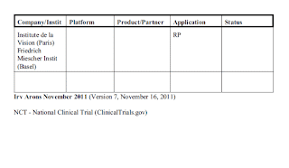
More treatments for retinitis pigmentosa are foreseeable in the coming decade, but genetic testing of RP patients is essential in order both to understand better the genetic associations of the disease and to direct efforts at developing treatments. However, even before implementing genetic testing, it is important to obtain, as much as possible, a complete family history in order to identify the inheritance pattern of the disease. The inheritance pattern has serious implications for the prognosis of the patient and is critical for genetic counseling for the patient and his or her family. RP
REFERENCES
1. Humphries P, Kenna P, Farrar J. On the molecular genetics of retinitis pigmentosa. Science. 1992;256:804-808.
2. McKusick VA, Mendelian Inheritance in Man: A Catalog of Human Genes and Genetic Disorders. Vol CD-ROM. 12th ed. Baltimore, MD; The Johns Hopkins University Press; 1998.
3. Berson EL, Gouras P, Hoff M. Temporal aspects of the electroretinogram. Arch. Opthalmol. 1969;81:207-214.
4. Bunker CH, Berson EL, Bromley WC, Hayes RP, Roderick TH. Prevalence of retinitis pigmentosa in Maine. Am J of Opthalmol. 1984;97:357-365.
5. Ayuso C, Garcia-Sandoval B, Najera C, Valverde D, Carballo M, Antinolo G. Retinitis pigmentosa in Spain. The Spanish Multicentric and Multidisciplinary Group for Research into Retinitis Pigmentosa. Clin Genet. 1995;48:120-122.
6. Lima LH, Cella W, Greenstein VC, et al. Structural assessment of hyperautofluorescent ring in patients with retinitis pigmentosa. Retina. 2009; 29:1025-1031.
7. Tsang SH, Vaclavik V, Bird AC, Robson AG, Holder GE. Novel phenotypic and genotypic findings in X-linked retinoschisis. Arch Ophthalmol. 2007; 125:259-267.
8. Tsui I, Chou CL, Palmer N, Lin CS, Tsang SH. Phenotype-genotype correlations in autosomal dominant retinitis pigmentosa caused by RHO, D190N. Curr Eye Res. 2008;33:1014-1022.
9. Zeiss CJ, Ray K, Acland GM, Aguirre GD. Mapping of X-linked progressive retinal atrophy (XLPRA), the canine homolog of retinitis pigmentosa 3 (RP3). Hum Mol Genet. 2000;9:531-537.
10. Tsang SH, Tsui I, Chou CL, et al. A novel mutation and phenotypes in phosphodiesterase 6 deficiency. Am J Ophthalmol. 2008;146:780-788.
11. Bainbridge JW, Smith AJ, Barker SS, et al. Effect of gene therapy on visual function in Leber's congenital amaurosis. N Engl J Med. 2008;358:2231-2239.
12. Maguire AM, Simonelli F, Pierce EA, et al. Safety and efficacy of gene transfer for Leber's congenital amaurosis. N Engl J Med. 2008;358:2240-2248.
13. Cideciyan AV, Aleman TS, Boye SL, et al. Human gene therapy for RPE65 isomerase deficiency activates the retinoid cycle of vision but with slow rod kinetics. Proc Natl Acad Sci U S A. 2008;105:15112-15117.
14. Gouras P, Kong J, Tsang SH. Retinal degeneration and RPE transplantation in Rpe65(-/-) mice. Invest Ophthalmol Vis Sci. Oct 2002;43:3307-3311.
Stephen H. Tsang, MD, PhD, is an ophthalmic geneticist and ERG attending at Columbia. Kyle Wolpert, BA, is a laboratory assistant at the Harkness Eye Institute of the Columbia University Medical Center. Neither author reports any financial interest in any products mentioned in this article. Dr. Tsang can be reached via e-mail at
dr.stemcells@gmail.com.